Sphenoid bone anatomy, parts and labeled diagram GetBodySmart
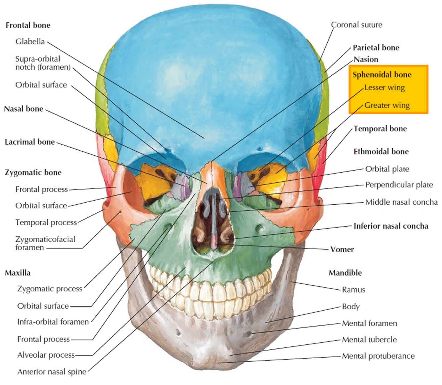
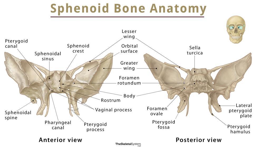
Normal Anatomy The sphenoid bone forms the central skull base and viewed anteriorly resembles a bird with its wings unfurled. It is a compound bone with a median body and paired lateral greater and lesser wings ( Fig. 3.1 ).
Sphenoid Bone Location, Function, Anatomy, & Labeled Diagram
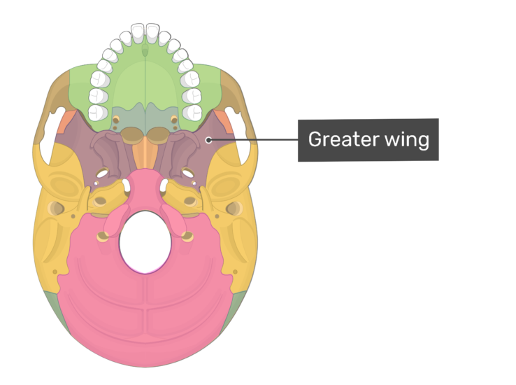
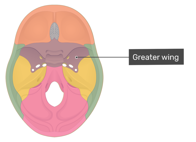
Greater Wings Emerging behind the lesser wings and also running to the sides are the two greater wings, which are also triangular and run lateral to the body. Their sides make up the infratemporal surfaces, which are convex in shape, and move backward and to the sides.
Sphenoid bone anatomy, function, parts & sphenoid bone fracture
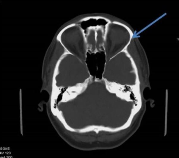
The greater wings of the sphenoid bone (GWS) are the components of the sphenoid bone that project laterally from the central body and form the anterior and medial portions of the floor of the middle cranial fossa. Many skull base foramina in the GWS transit important neurovascular structures.

Greater Wing Of Sphenoid slidesharedocs
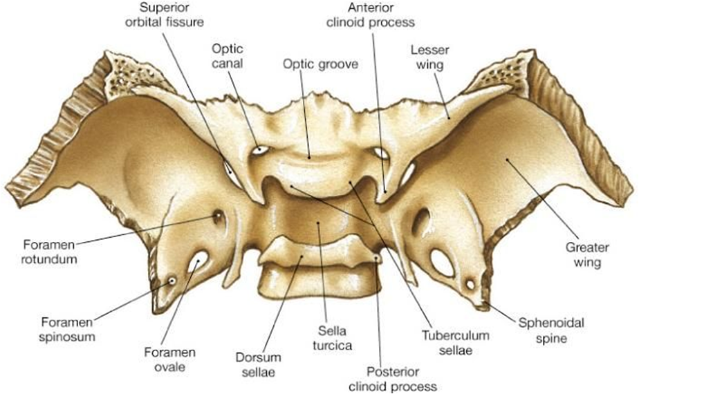
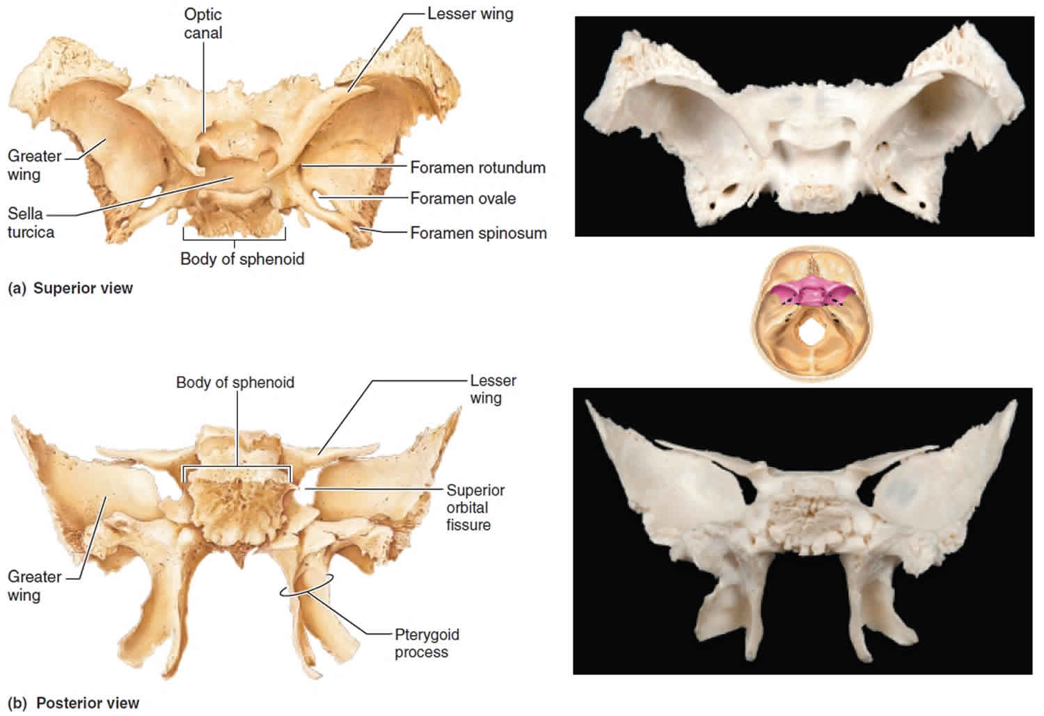
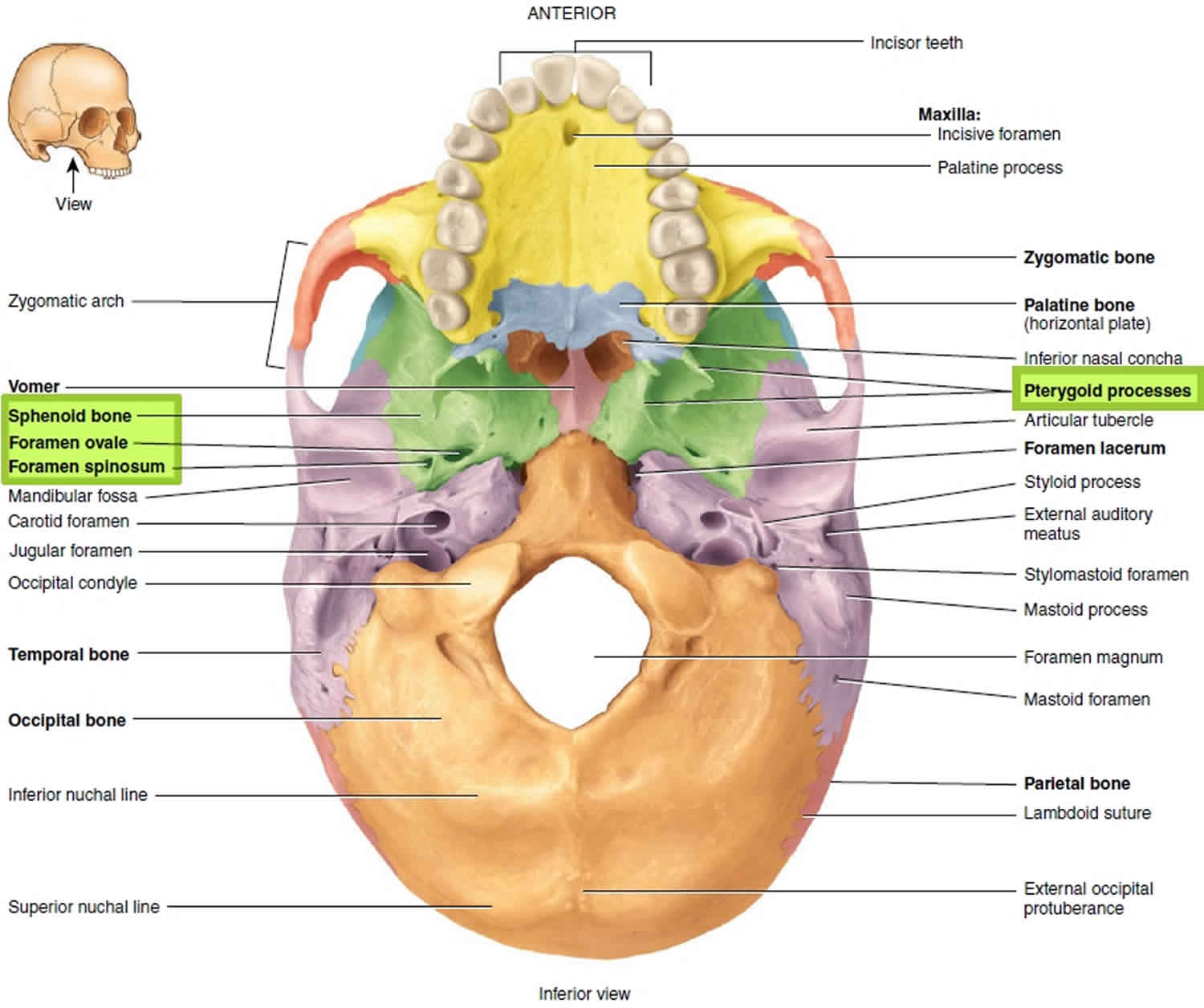
greater wing lesser wing pterygoid process and plates Articulations The sphenoid bone articulates with twelve bones: unpaired bones include: frontal, ethmoid, vomer, and occipital paired bones include: zygomatic, parietal, temporal, and palatine. Fissures, foramina, grooves and canals The sphenoid bone includes: optic canal superior orbital fissure
Anterior View of Sphenoid Bone Neuroanatomy The Neurosurgical Atlas
Functions It forms the base and lateral sides of the skull in combination with the orbital floor. Anatomy: Parts and Structure of the Sphenoid Bone The sphenoid bone consists of a central body, with two lateral paired wings on either side - the lesser and greater wings - and two pterygoid processes.

Treating TMJ issues the role of the sphenoid bone MaryAnn Reynolds
Sphenoid wing meningiomas are slow growing tumors that originate from outer arachnoid meningeal epithelial cells. They are the most common tumor of the intracranial space to spread to the orbit. [1] [2] Contents 1 Disease Entity 1.1 Incidence 1.2 Etiology 1.3 Systemic Associations 1.4 General Pathology and Classification 2 Diagnosis

[PDF] Medial sphenoid wing meningioma. Semantic Scholar
The greater wings of the sphenoid bone (GWS) comprise the com-ponents of the sphenoid bone that make up most of the posterior orbital wall and form the anterior and medial parts of the floor of the middle cranial fossa. Many important skull base foramina, which transmit vital neurovascular structures, are present in these paired wings on either.

Sphenoid bone anatomy, function, parts & sphenoid bone fracture
Synonyms: none The sphenoid bone is one of the most complex bones of the human body. Due to its shape, it is also referred to as the 'wasp bone'. It makes up most of the middle part of the base of the skull and contributes to the floor of the middle cranial fossa of the skull.
Greater Wing Sphenoid CT Tutor
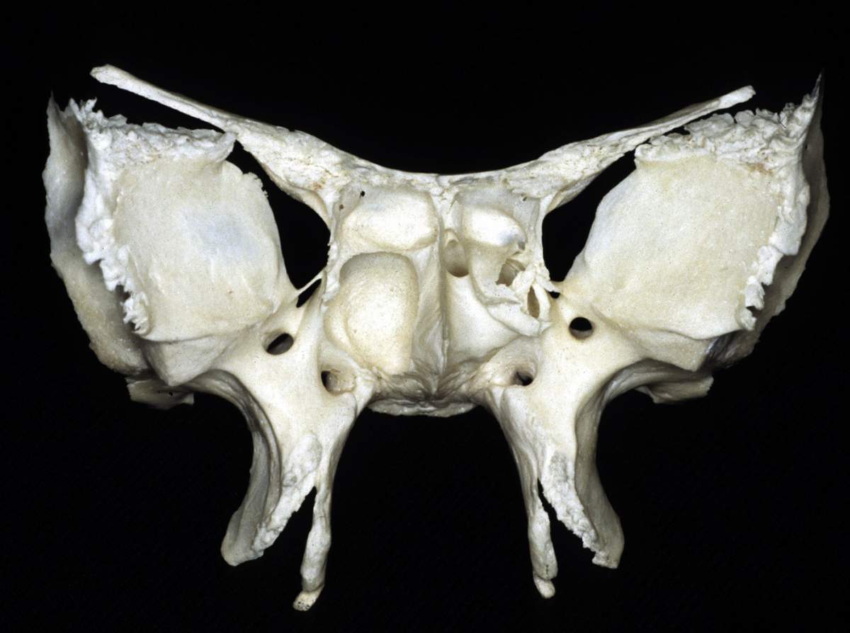
The greater wing of the sphenoid is a bony projection arising from both sides of the sphenoid body forming a part of the floor of the middle cranial fossa. Jakob Benignus Winslow coined the term foramen spinosum because of the foramen's location in the spinous process of the greater wing of the sphenoid. [1] Go to: Structure and Function
😊 Sphenoid bone location. Sphenoid Sinus Anatomy, Diagram & Location
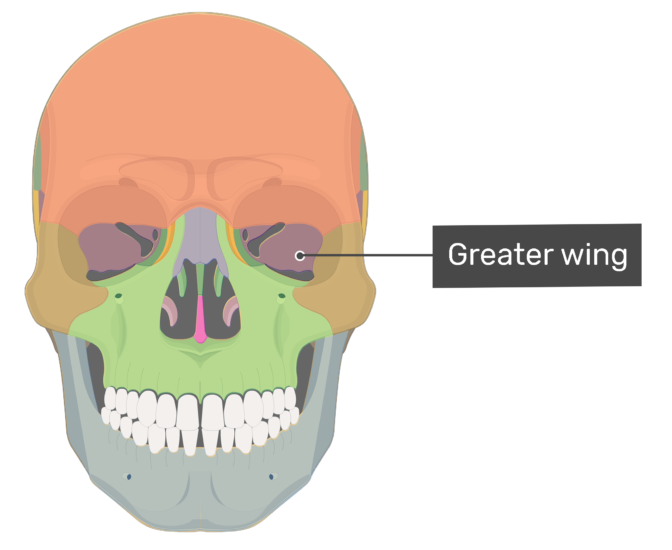
The greater wing of the sphenoid bone can be seen in the below image as the oval shape in the center of the red rectangle. Site of the pterion. The sphenoid bone body sits behind the nasal cavity with various protrusions that play important roles in the construction of other bony structures.
easyhumanatomy Sphenoid easy and brief discussion
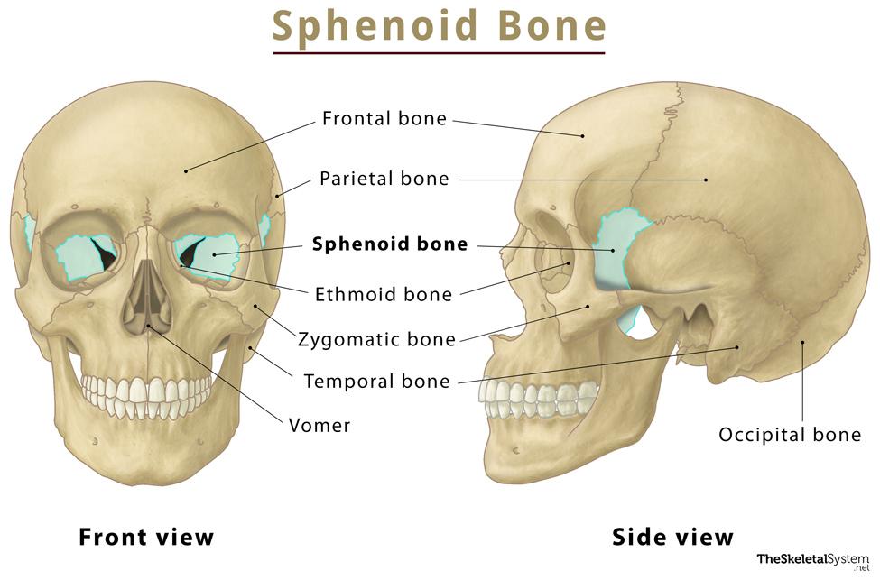
The sphenoid is just one of the twenty-two bones that form the skull and essentially helps to connect the neurocranium to the facial skeleton. It is a single bone in the midline of the cranial cavity situated posterior to the frontal bone but anterior to the occipital. Its name derives from the Greek 'sphenoeides,' which means wedge-shaped.
Sphenoid bone anatomy, function, parts & sphenoid bone fracture
The sphenoid bone (also called the sphenoid, sphenoidal bone, Latin: os sphenoidale) is a single bone that lies in the base of the skull between the frontal, temporal and occipital bones. The sphenoid bone has a central body, and paired greater and lesser wings that extend laterally from the body of the sphenoid.
Sphenoid bone anatomy, parts and labeled diagram GetBodySmart
The greater wing of the sphenoid bone also forms the anterior margin of the foramen lacerum, which is filled with cartilage during life. Anterior to the greater wing of the sphenoid is the lesser wing of the sphenoid bone. This shelf of bone forms the posterior part of the anterior cranial fossa and extends laterally, and articulates.
Sphenoid Bone Location, Function, Anatomy, & Labeled Diagram
two greater wings on the lateral side of the body and two lesser wings from the anterior side. Pterygoid processes of the sphenoides, directed downwards from the junction of the body and the greater wings. [4] Two sphenoidal conchae are situated at the anterior and inferior part of the body. Intrinsic ligaments of the sphenoid
Sphenoid Bone
Anatomical Structure The sphenoid bone is said to be ' butterfly-shaped '. It consists of a body, paired greater wings and lesser wings, and two pterygoid processes. Body The body lies at the centre of the sphenoid bone and is almost completely cubiodal in shape.
Sphenoid bone anatomy, function, parts & sphenoid bone fracture
greater wing of sphenoid (bone): [TA] strong squamous processes extending in a broad superolateral curve from the body of the sphenoid bone. The greater wing presents these surfaces (facies): 1) cerebral surface: forms anterior third of the floor of the lateral portion of the middle cranial fossa; 2) temporal surface: forms the deepest portion.